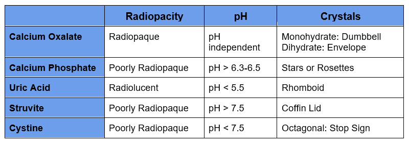
Table of Contents
Kidney stones are common and the incidence is increasing. Knowledge of management and prevention is imperative.
This article will approach diagnosis. Not diagnosing that a patient has a kidney stone, but diagnosing the type of stone and assessing for secondary causes in patients with recurrent nephrolithiasis.
Types of Kidney Stones
The five most common types of kidney stones are
- Calcium Oxalate
- Uric Acid
- Calcium Phosphate
- Struvite
- Cystine
Rare causes
- Xanthine
- 2,8-dihydroxyadenine
- Drug/Medication Stones
Determining Stone Composition
Stone Analysis
The best way to determine stone composition is a stone analysis report. The analysis will often give the chemical and/or mineralogic name of the stone. For each stone type there are different chemical/mineralogic subtypes. Knowledge of these is necessary to know what type of stone you are dealing with.
Calcium oxalate
- Calcium oxalate monohydrate: Whewellite
- Calcium oxalate dihydrate: Weddellite
This is by far the most common stone. Most stones have a combination of these two subtypes, and may even have three components also containing a calcium phosphate component.
- If the stone is composed solely of the monohydrate or dihydrate forms an underlying congenital or hereditary condition is more likely.
- Calcium oxalate monohydrate
- Harder composition. More difficult to fragment with lithotripsy
- More common with hyperoxaluria.
Calcium phosphate
- Carbonate apatite
- Hydroxyapatite
- Calcium hydrogen phosphate: Brushite
There are multiple forms of calcium phosphate. They are often combined with calcium oxalate. If the major component is a form of calcium phosphate evaluate for secondary causes of this stone type
Uric acid
- Uric Acid
- Uric Acid dihydrate
- Ammonium acid urate
- Sodium urate
Struvite
- Magnesium ammonium phosphate: Struvite
These are infection stones from urea splitting organisms
Cystine
These stones form with cystinuria.
If Stone Analysis Not Available
Every attempt should be made to analyze the stone. However, if not available there are clinical clues that may help.
- Is the stone radiopaque or radiolucent?
- What is the urine pH?
- Are there crystals on urinalysis?
This refers to if the stone is visible on a KUB X Ray. All types of stones can be seen on CT which has the highest sensitivity (97%). All stone types can also be seen on ultrasound, although there is a poorer sensitivity (50%). Only radiopaque stones can be seen on X Ray although there is also a poor sensitivity.
Uric acid stones form in an acidic pH < 5.5. Struvite stones form in an alkaline urine pH 7.5. Calcium phosphate stones also precipitate in an alkaline pH, albeit more modest > 6.3-6.5. Calcium oxalate stones precipitation is pH independent.
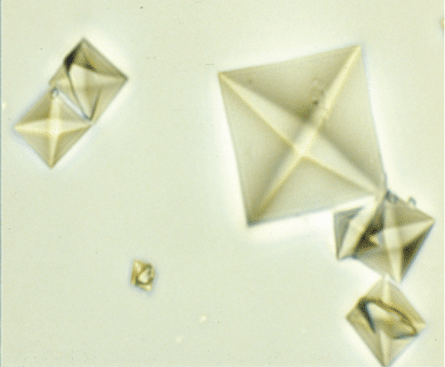
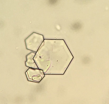
Each stone has different crystals that can be seen under microscopy. With the exception of cystine, crystals can normally form ex vivo at the lower temperatures outside the body. There are 2 types of crystals that are characteristic and I can easily see. These are calcium oxalate dihydrate which are envelope shaped and cystine crystals which are octagonal and look like little stop signs.
Crystals
Calcium Oxalate Dihydrate
Cystine
Causes of Kidney Stones
Each type of stone may have underlying secondary causes, although many are idiopathic and/or attributed to dietary factors. Know these to diagnose potential underlying conditions
Calcium Oxalate:
- Hypercalciuria
- Hypercalcemia: Any cause of hypercalcemia (with the exception of thiazide diuretics) can cause hypercalciuria which in turn is a risk factor for stones. These include:
- Primary hyperparathyroidism. Although thiazides decrease urinary calcium, they may unmask primary hyperparathyroidism, be aware of this
- Sarcoidosis
- Milk – Alkali syndrome
- Vitamin D excess
- Supratherapeutic Vitamin D levels
- Calcitriol
- Hypercalcemia: Any cause of hypercalcemia (with the exception of thiazide diuretics) can cause hypercalciuria which in turn is a risk factor for stones. These include:
It is my belief that Vitamin D2 or Vitamin D3 without elevated levels does not cause hypercalciuria and is safe to use in patients with nephrolithiasis. Calcitriol can cause hypercalciuria and should be avoided.
Vitamin D2? Vitamin D3? 25 Vitamin D? 1,25 Vitamin D?
Idiopathic hypercalciuria. With improved genetic analysis, genetic causes are becoming more common realized causes of “idiopathic’
-
- Dent Disease (X linked)
- Lowe Syndrome (X Linked)
- Others
- Hyperoxaluria
- Primary Hyperoxaluria
- Enteric Hyperoxaluria (Bariatric surgery, bowel disease)
How Does Bariatric Surgery Cause Kidney Stones? @BCNephro – YouTube
Calcium Phosphate:
- Hypercalciuria (as above)
- Particularly primary hyperparathyroidism which may also be associated with an alkaline urine pH
- Alkaline urine pH
- Distal RTA. In addition to a high urine pH distal RTA can cause hypercalciuria and hypocitraturia. A incomplete distal RTA (associated normal serum bicarbonate, alkaline urine pH, and hypocitraturia) or drug induced RTA (Topiramate) are causes of calcium phosphate nephrolithiasis.
Uric acid:
- Alkaline urine pH
- Unlike gout there may not be hyperuricemia. The main risk factor is a low urine pH. An acidic urine pH occurs with a high animal protein diet.
Struvite:
- Urine infections
- Urea splitting organisms (Proteus; Klebsiella)
Cystine
- Cystinuria
- Autosomal Recessive condition.
Summary
In a patient with recurrent nephrolithiasis stone compositions should be determined. This is best done by kidney stone analysis. If stone analysis is not available, radiopacity, urine pH, and the presence of crystals can provide clinical clues as to the stone type.
Secondary causes of calcium stones include most causes of hypercalcemia, particularly primary hyperparathyroidism. Enteric hyperoxaluria is a cause of calcium oxalate stones and causes of elevated pH such as distal RTA which may be medication induced from topiramate is associated with calcium phosphate stones.
The main risk factor for uric acid stones is an acidic urine pH which often occurs with a high animal protein diet. Alkaline urine pH from urea splitting urinary infections is the cause of struvite stones.

